神经细胞能再生的技术(神经再生研究新突破)
来自北京航空航天大学、首都师范大学、上海交通大学等机构的研究人员展开合作,在脊髓损伤再生研究中取得突破性的成果。两任职于北京航空航天大学及首都师范大学的李晓光(Xiaoguang Li)研究员,同济大学医学院和加州大学洛杉矶分校的孙毅(Yi E. Sun)教授,以及李晓光课题组的杨朝阳(Zhaoyang Yang)博士是这篇论文的共同通讯作者。
Transcriptome analyses reveal molecular mechanisms underlying functional recovery after spinal cord injury.October 12, 2015, doi: 10.1073/pnas.1510176112 PNAS October 12, 2015
由于恶劣的中枢神经系统(CNS)损伤环境以及CNS神经元较弱的内在再生能力, CNS中的轴突再生极具挑战性,因此脊髓损伤(SCI)被认为无法治愈。
在这篇文章中研究人员发现,载有神经营养因子-3(NT3)的壳聚糖可 提供良好的环境,促进大鼠神经生长、新神经发生及完全切断的脊髓恢复功能。为了了解其分子机制,研究人员在脊髓损伤后90天时间内对损伤位点的脊髓段,以 及损伤吻侧和尾侧区域进行了一系列全面的转录组分析。利用加权基因共表达网络分析(Weighted gene co-expression network analysis, WGCNA),他们确立了在脊髓损伤后不同时间响应各种病理事件的基因模块/程序。客观测量基因模块表达还揭示出新神经发生和血管发生增加,炎症反应减少 是NT3-chitosan影响再生的关键。
NT3-chitosan elicits robust endogenous neurogenesis to enable functional recovery after spinal cord injury.October 12, 2015, doi: 10.1073/pnas.1510194112 PNAS October 12, 2015
成年哺乳动物中枢神经系统中的神经干细胞(NSCs)通过适当激活、分化和成熟,建立起新神经网络,整合到受损神经回路中修复功能,是神经再生的关键。然而,中枢神经系统损伤微环境往往是抑制及炎症性的,限制了活化的神经干细胞分化为神经元,形成新神经回路的能力。
在这篇文章中,研究人员报告称当将偶联NT3的壳聚糖材料插入到完全切断和切除的大鼠胸部脊髓5-mm缺口中时,可引起受损脊髓中內源NSCs有力 地活化。通过缓慢释放NT3,这一生物材料吸引NSCs迁移到了损伤区域,分化为神经元,形成了功能性神经网络,其与切断的上行和下行轴突相互连接,导致 了感觉与运动行为恢复。
这项研究提出了,促进内源性神经发生是脊髓损伤一种新治疗策略。

Proc Natl Acad Sci U S A. 2015 Oct 27; 112(43): 13360–13365.Published online 2015 Oct 12. doi: 10.1073/pnas.1510176112PMCID: PMC4629389Neuroscience
Transcriptome analyses reveal molecular mechanisms underlying functional recovery after spinal cord injury
Hongmei Duan,a,1 Weihong Ge,b,1 Aifeng Zhang,c,1 Yue Xi,a,1 Zhihua Chen,d,1 Dandan Luo,e Yin Cheng,b Kevin S. Fan,f Steve Horvath,g Michael V. Sofroniew,h Liming Cheng,i Zhaoyang Yang,a,j,2 Yi E. Sun,e,b,2 and Xiaoguang Lia,j,2Author information ► Copyright and License information ►See "NT3-chitosan elicits robust endogenous neurogenesis to enable functional recovery after spinal cord injury" in volume 112 on page 13354.This article has been cited by other articles in PMC.Go to:
SIGNIFICANCE
In this study, we used gene expression analyses to unveil mechanisms underlying NT3-chitosan–induced spinal cord regeneration. Using a powerful bioinformatics tool known as weighted gene coexpression network analysis, we have established gene modules and programs representing various events at different times after spinal cord injury (SCI) and also demonstrated that enhanced new neurogenesis and vascularization, as well as reduced inflammatory responses, are keys to conferring the effect of NT3-chitosan on regeneration. The objectivity of this approach and the use of big data processing have opened a new pathway in SCI research. Such quantitative, objective, and sensitive measures could provide a standardized approach in the future to reveal mechanistic insight into various potential interventions for SCI repair.
Keywords:NT3, chitosan, WGCNA, spinal cord injury, transcriptomeGo to:
ABSTRACT
Spinal cord injury (SCI) is considered incurable because axonal regeneration in the central nervous system (CNS) is extremely challenging, due to harsh CNS injury environment and weak intrinsic regeneration capability of CNS neurons. We discovered that neurotrophin-3 (NT3)-loaded chitosan provided an excellent microenvironment to facilitate nerve growth, new neurogenesis, and functional recovery of completely transected spinal cord in rats. To acquire mechanistic insight, we conducted a series of comprehensive transcriptome analyses of spinal cord segments at the lesion site, as well as regions immediately rostral and caudal to the lesion, over a period of 90 days after SCI. Using weighted gene coexpression network analysis (WGCNA), we established gene modules/programs corresponding to various pathological events at different times after SCI. These objective measures of gene module expression also revealed that enhanced new neurogenesis and angiogenesis, and reduced inflammatory responses were keys to conferring the effect of NT3-chitosan on regeneration.
Spinal cord injury (SCI) is a debilitating medical condition that often leads to permanent impairment of sensory and motor functions. SCI is considered almost incurable because axons in the central nervous system (CNS), unlike those in the peripheral nervous system (PNS), are believed not to regenerate. The innate ability of mature CNS neurons to regenerate is much weaker than that of PNS neurons (1). In addition, myelin debris in the injured CNS is more inhibitory toward axonal growth compared to that in the PNS (2). Moreover, the mode of immune cell infiltration and microglia activation are different in CNS versus PNS, resulting in a different cellular microenvironment, which crucially influences the outcome, i.e., PNS axons regenerate, while CNS axons do not (3).
Over the years, SCI research has focused on ways to promote the long-distance growth of CNS motor axons, mainly by neutralizing inhibitory myelin components and/or changing the neuronal intrinsic program to enable better regeneration (4). Unfortunately, however, although numerous studies have been carried out following this line of strategy, no major breakthroughs translatable to therapy have been achieved. In recent years, efforts toward promoting long distance axonal growth have been complemented with alternative approaches aimed at using exogenous stem cells to generate local new neurons that form nascent relay neural networks to pass ascending and descending neurotransmission signals with or without long-distance axonal growth (5–7).
SCI is a complex medical condition. The primary lesion includes the physical traumatic wounding of both white and gray matter, breakdown of the vasculature system, and acute immune reactions, which is followed by secondary lesions, such as demyelination, additional immune cell infiltration, inflammation, glial scar formation, impaired neurotransmission, and neuronal apoptosis (8). Secondary lesions are intermingled with intrinsic repair processes, including remyelination, reestablishment of the vasculature system, reactivation of presumptive endogenous neural stem/progenitor cells (NSCs), and axonal sprouting and growth (9). In the past, these various pathological events have been studied in an isolated or incoherent manner without assembling the factors into a framework to provide a holistic view. One major challenge in the field of SCI research is the huge variability caused not only by different lesion models and animal strains, but also by even subtle changes in the exact lesion operations. Moreover, most analyses for evaluating neural repair/regeneration have been anatomic, behavioral, and sometimes electrophysiological, including extracellular recordings (1, 4, 10, 11). These analyses are relatively low in resolution, and particularly the behavioral assays can be highly variable and subjective to errors in human judgment. As a result, the SCI research field has suffered from substantial inconsistency and irreproducibility of various reported findings, results, and conclusions.
In addition to double-blind designs of behavioral assays proposed as necessary, the development of more objective, quantitative, and uniform measures of various perspectives of pathological events in the injured spinal cord will be hugely beneficial to SCI research efforts. A cell can be generally defined by the set of genes that it expresses. Therefore the transcriptome reflects not only the identity of the cell, but also the physiological/pathological state of that cell. Presumably, within the injured spinal cord at different time points postinjury, various pathological cellular events could be reflected by transcription programs of injured spinal cord segments (8). Unfortunately, the lack of powerful bioinformatics data processing capability had confounded previous attempts of using transcriptome analyses to study SCI (8); however, a recently developed novel analytical approach, weighted gene-coexpression network analysis (WGCNA), now allows the identification of cores of gene networks based on a pairwise Pearson correlation matrix of all expressed genes across samples (12, 13). Genes with similar expression patterns across all samples will innately cluster together. These clustered cores, or modules, often represent genes involved in the execution of particular biological functions, including vasculature development, apoptosis, neuronal differentiation, and synaptic transmission, etc. Moreover, gene functions in particular cell types sometimes also cluster together, and thus some modules could be specific for oligodendrocytes/myelination, immune cells/defense response, neurons/synaptic transmission, and so forth. These events actually represent cell type-specific functions. The WGCNA method allows dissection of various ingredients (i.e., various pathological events) in the “soup” of an injured spinal cord via transcriptome analyses (14).
In the present study, we used WGCNA to establish gene module series representing major pathological events in a temporally and spatially specific manner following complete transection and extraction of the thoracic spinal cord, which can serve as objective outcome measurements. Using these measures, we further identified critical mechanisms underlying robust functional recovery by neurotrophin-3 (NT3)-chitosan after SCI.
Go to:
RESULTS
Experimental Design for Spinal Cord Transcriptome Analysis Post-SCI.
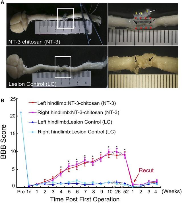
In previous studies, we found that NT3-loaded chitosan biomaterial had a profound effect on promoting spinal cord regeneration as well as motor and sensory functional recovery (Fig. S1). To explore the molecular mechanisms by which NT3-chitosan facilitated spinal cord regeneration, we carried out extensive transcriptome analyses. Wistar female rats (200–220 g) were used for all experiments. Complete transaction of the thoracic spinal cord at T7-8 was performed, with a 5-mm segment of the cord removed. The emptied space was either left alone [lesion control (LC)] or refilled with a 5-mm chitosan tube either loaded with NT3 or not loaded with NT3 [empty tube (ET)] before the dura was resutured. The success of the experiment was evaluated by parallel anatomical analysis at 90 d postsurgery demonstrating the growth of nerve-like bridging tissues within the NT3-chitosan tube, as well as behavioral analyses with a double-blind design indicating substantial functional recovery in the NT3-chitosan treatment group (Fig. S1).

Fig. S1.NT3-chitosan promotes nerve regeneration and motor functional recovery after complete SCI in rats. (A) Anatomic images (dorsal view) of regenerating nerve tissue (black arrows) in NT3-chitosan tube (NT3) or scar tissue (black arrows) in LC samples at ...
Animals were euthanized at 1, 3, 10, 20, 30, 60, and 90 d postsurgery, and 5-mm segments of the spinal cord at the lesion site, as well as immediately caudal or rostral to the lesion segment, were collected and labeled with “R” for rostral, “C” for caudal, and “M” for lesion site (Fig. 1A). Spinal cord segments from four animals together were needed to provide sufficient RNA for microarray analyses as a single sample; thus, a total of 12 animals were euthanized to provide three samples at each time point. Quality mRNA was extracted, reverse-transcribed, and subjected to Affymetrix GeneChip (Rat Genome 230) 2.0 array analysis. Three uninjured spinal cord control samples corresponding to the lesion segment (5 mm) at T7-8 from 12 animals were included. Five reference RNA samples included in the microarray kit were used to control for a potential microarray batch effect.

Fig. 1.Sample clustering revealed temporal and spatial changes following SCI with or without NT3-chitosan treatment. (A) A schema describing anatomic positions of spinal cord segments for the LC and NT3 samples used for transcriptome analyses. Empty chitosan ...
Region-Specific Changes Following SCI Revealed by Sample Clustering.
Unbiased hierarchical clustering of the complete dataset (167 samples from 668 animals) based on Pearson’s correlation coefficient of transcriptomes among pairs of samples in all combinations divided the samples into two major groups. All M region samples formed one cluster, and all R and C samples together formed the other large cluster (Fig. 1B). Three uninjured normal spinal cord samples clustered together (Fig. 1B, blue circle, white arrow) and were more closely correlated with R and C samples than with M samples, suggesting that more dramatic injury repair and tissue reconstitution events occurred within the M regions, whereas smaller changes occurred in R and C regions (Fig. 1B). In the figure, the white-framed stripes present information on correlations between the uninjured group and the rest of the samples; the darker the color, the better the correlation.
Sample clustering provided more information. For example, within the M region, NT3-chitosan–treated samples (labeled in green in Fig. 1B) tended to cluster together, and these samples correlated better with uninjured control samples than LC samples (labeled in red in Fig. 1B). Moreover, samples from the R and C regions that correlated best with uninjured control samples tended to be NT3-chitosan–treated samples and samples obtained at both early (day 1) and late (day 90) time points (Fig. 1B), suggesting that NT3-chitosan treatment had beneficial effects soon after injury and resulted in better recovery in the long run. Two-dimensional principal component analysis (PCA) of the whole data matrix further demonstrated that transcriptome differences caused by regional differences were greater than those caused by time differences, and those caused by time differences were greater than those caused by treatment differences (Fig. 1C). This suggests that (i) the M region underwent the most dramatic biological changes; (ii) different biological/pathological events occurred at different time points postinjury, which could be reflected in the transcriptome; and (iii) NT3-chitosan had beneficial effects, resulting in closer resemblance to uninjured cord, as indicated by the fact that most filled dots (NT3) were closer to uninjured samples compared with other treatment samples in the same region and at the same time (Fig. 1C).
Using WGCNA to Establish Gene Modules Underlying Pathological Events Post-SCI.
Given that the M region underwent drastic tissue reconstitution starting with an empty space for both LC and NT3 groups (ET group was omitted as no tissue growth in the M region was observed), we expected that biological/pathological events occurring in R and C regions would be different from or less robust than those in the M regions, and thus should be analyzed separately. We first subjected uninjured/normal samples and all LC samples in R and C regions to WGCNA, with an emphasis on genes that underwent lesion-induced changes (a total of 7,500 annotated genes were included), which should digitally represent pathological cellular events in the lesion area after complete transection of the spinal cord. WGCNA identified 10 gene clusters/modules (designated as C1–C10) of coexpressed genes (Fig. 2 A and B).

Fig. 2.WGCNA of LC samples in R and C regions identified gene modules underlying pathological events at different time points post-SCI. (A) Hierarchical cluster dendrogram of LC samples in R and C regions showing coexpression modules identified using WGCNA on ...
Gene Ontology (GO) analysis of these 10 modules revealed several key biological processes that occurred in a temporally specific manner postinjury (Fig. 2B and Dataset S1). GO terms of each module revealed that synaptic transmission-related genes (C1), including neurotransmitter receptors, synaptic proteins, and ion channels (Chrna7, Grm1, Syn2, and Kcnc1), as well as axon/dendrite-related genes and calcium-signaling genes (Amigo, Stxbp1, Map1b, Map2, Atp2b2, Camk2b, and Calm3), underwent dramatic down-regulation immediately after injury and remained low even at day 90 postsurgery, indicative of long-term impairment. Genes involved in sterol/cholesterol biosynthesis, tRNA metabolism, and ubiquitin-dependent protein degradation (C2) exhibited reduced expression with a slightly slower kinetics compared to C1, and also remained low throughout the 90-d period. In contrast, expression of genes involved in immune response, defense, inflammation, and cell death (C10:Tlr4, Tgfb1, Vav1, C1qc, C1qb, and Casp1) rose quickly after injury and remained high throughout the 90-d period, whereas expression of genes in another immune response (innate and adaptive) and wounding response module (C8: Ctsh, Ctsd, Ctsa, Hexa, Cd86, Tnfsf4 Igf1, C2, C4, B2m, and Tgfbr) increased slightly slower than those in C10, yet remained high for at least 90 d postinjury. In C3, a module related to neuronal differentiation and projection (Gja1, Lingo1, Pax6, Epha4, Ntrk2, Rpe65, and CamK2g), astrocyte and oligodendrocyte development (Aldh1l1, Cd24, Cnp, Omg, Id4, and S100b), and oxidation reduction (Dhtkd1, Bcan, Glud1, Prodh, and Cyp2d4v1), gene expression decreased immediately after injury but then reversed relatively quickly during the first 10 d post-SCI, and continued to slowly increase, reaching almost normal (uninjured) levels by day 90. This suggests that demyelination-remyelination and astrogliosis were dynamically regulated during the first 10-d period postinjury, and gradually diminished after 10 d. Likewise, C6 and C7, two modules related to cell cycle, apoptosis (Fbl, Ddx39, and Thoc4), and inflammation (Il6, jmjd6, and Cxcl1/2/7) rose quickly postinjury and then dropped rapidly over the first 10 d, and thereafter continued to gradually revert back to close to normal (uninjured) levels. C4 and C9, two modules related to metabolism of fatty acid, glycogen, mRNA and energy reserve, and vasculature development, also were dynamically regulated during the first 10 d and continued to increase slowly over the 90-d period. The unique module C5, involved in antigen processing and coenzyme metabolism, exhibited an “up-and-down” wavy time course. The biological significance of such changes remains to be elucidated.
Of note, as indicated by differential gene expression analyses, in LC samples, overall gene expression changes were not statistically significantly different in R regions versus C regions; only 0.2% of genes were differentially expressed in the two regions (P < 0.01). In contrast, in ET samples, 7% of the genes (i.e., 1,400 of 18,000) were differentially expressed in R versus C regions. These 1,400 genes were associated with cell adhesion, neuron projection, vasculature development, cell–cell signaling, and regulation of neurologic system processes, which appeared to be regulated more robustly in R regions. Considering that ET formed a physical barrier between R and C regions, since nothing grew in the chitosan-only tube, differential gene expression in R versus C regions suggested different injury signals were originated in the two regions. Under LC conditions, cellular components may shuttle between R and C regions, passing information back and forth to trigger similar injury responses, whereas such shuttling should be blocked in the ET group, resulting in different gene regulation in R versus C regions. In NT3 samples, approximately 518 of 18,000 genes were differentially expressed in R versus C regions, suggesting that NT3-chitosan connects the two regions, because cells did migrate into the tube. Moreover, NT3 appeared to elicit more robust changes in vasculature development, neuron development, and synaptic transmission in R regions than in C regions.
Taken together, our data indicate that WGCNA is capable of providing a digitized image of pathological events after complete spinal cord transection injury. Based on this information, outcome measurements could be established to evaluate various putative therapeutic interventions for SCI repair in a temporally and spatially specific manner.
NT3-Chitosan–Induced Regeneration Mediated by Antiinflammation and New Neurogenesis Following SCI.
From PCA, it was apparent that gene regulation was more dynamic in the M region than in R and C regions, consistent with the fact that tissue reconstitution occurred in the M region. To strengthen the investigation to identify key cellular and molecular programs influenced primarily by NT3-chitosan, we carried out WGCNA on M region samples with noninjury controls. This analysis identified 19 modules (Fig. 3A), 7 of which showed statistically significant differences upon NT3-chitosan treatment (Fig. 3B). Analysis of the module–trait relationship identified five modules (Cm1, Cm2, Cm3, Cm4, and Cm5) as positively correlated with NT3-chitosan treatment and two modules (Cm6 and Cm7) as negatively correlated with NT3-chitosan treatment (P < 0.05) (Fig. 3B).

Fig. 3.WGCNA of uninjured and all M region injury samples uncovered molecular mechanisms underlying the proregeneration function of NT3-chitosan. (A) Hierarchical cluster dendrogram using WGCNA analyzing uninjured samples (N) and samples in the M regions. A ...
GO and hub gene network analyses, shown in Fig. S2 and Dataset S2, indicated that Cm1 and Cm2 were related to neurogenesis; Cm4 was related to muscle contraction; Cm3 was related to pain and temperature sensory processing; Cm5 was related to angiogenesis, response to oxygen levels, and nervous system development; Cm6 was related to immune response; and Cm7 was related to response to wounding, stress, and inflammation. The averaged gene expression of each module further indicated that NT3-chitosan enhanced vascularization (blood supply), which was required for proper tissue repair. NT3-chitosan also clearly suppressed inflammatory responses (Cm6 and Cm7), which were classically considered key to attenuating secondary lesions after SCI. Therefore, the antiinflammatory effect elicited by NT3-chitosan also could contribute to the better regeneration outcome.

Fig. S2.Hub gene networks of seven modules significantly influenced by NT3-chitosan in M region injury samples.
One exciting finding was the enhanced expression of modules Cm1 and Cm2 (related to neurogenesis and neurotransmission) in response to NT3-chitosan. Moreover, Cm3 and Cm5 also contained many neurogenesis genes, including Dlx5, Isl2, Trpv1, Chrna3, Ntrk1, Ngfr, Agrn, GDNF receptors, Nrep, Smo, and Gata2. Although NT3-chitosan enhanced gene expression in Cm1 and Cm 2 and in Cm3 and Cm5, these four modules behaved differently. Cm1/2 represented genes related to neuronal differentiation, which were expressed at high levels in uninjured/normal spinal cord, whereas Cm3/5 represented neuronal differentiation genes, which were not highly expressed in normal adult spinal cord but were elicited de novo by NT3-chitosan. Interestingly, a muscle contraction module (Cm4) also was heavily induced by NT3-chitosan in the M region. This finding was unexpected and merited further investigation to determine the contribution of this module to better functional recovery after SCI. Expression levels of candidate genes from Cm1 (Dcx and Snap25), Cm2 (Ntrk3), and Cm5 (Dlx5) across all samples (all regions and all times) demonstrated robust regulation of these genes by NT3-chitosan in the M region compared with R and C regions (Fig. 3D). Expression plots for additional genes involved in NSC differentiation are shown in Fig. S3.

Fig. S3.Averaged expression of some representative genes related to neuronal and oligodendroglial differentiation across all samples. Note that NT3-chitosan promoted not only neuronal differentiation, but also oligodendroglial differentiation, in the M region ...
Taken together, our data from transcriptome WGCNA indicated that NT3-chitosan served primarily to enhance vascularization and suppress inflammatory immune responses, establishing an optimal microenvironment to support endogenous NSCs to differentiate into new neurons, which subsequently formed nascent local neural networks to participate in regeneration after SCI (15).
Biological Confirmation.
To assess whether NT3-chitosan indeed served as an antiinflammatory agent in our experiments, we stained injured spinal cord with NT3-chitosan treatment or no treatment using an anti-CD45 antibody, which labeled all infiltrating leukocytes (Fig. 4 A and B), and found reduced leukocyte infiltration in the NT3-chitosan group. To examine whether it was the chitosan material or NT3 per se, or the two together that elicited the maximum anti-inflammatory effect, we measured inflammatory cytokines from a mouse macrophage RAW cell line cocultured with lipopolysaccharide (LPS; positive control), chitosan, soluble NT3, and NT3-conjugated chitosan (NT3-chitosan). Our results demonstrated that at 1 d and 3 d after treatment, IL-1α/β, IL-6, NF-kb, and TGF-β expression and secretion were increased with LPS treatment, little changed with chitosan only or soluble NT3 treatment, but significantly decreased with NT3-chitosan treatment, suggesting that NT3-loaded chitosan could significantly suppress inflammatory cytokine production from RAW cells (Fig. 4C).
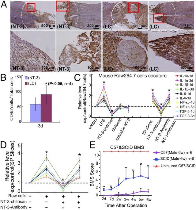
Fig. 4.NT3 chitosan creates a reduced inflammatory environment, which is beneficial for functional recovery post-SCI. (A and B) CD45 staining to label inflammatory leukocytes in injured rat spinal cord sections for both NT3 and LC groups and quantitative analyses ...
Moreover, we found that when RAW cells were cocultured with rat spinal cord slices, the aforementioned inflammatory cytokine secretion was increased as well, which could be suppressed by NT3-chisosan, and that NT3 antibody could block the effect of NT3-chitosan. Of note, we observed the same phenomenon when measuring mouse-specific cytokines (generated from RAW cells) or rat-specific cytokines (generated from rat spinal cord slices) (Fig. 4 C and D and Fig. S4).

Fig. S4.(A and B) ELISA demonstrating that NT3-chitosan suppressed the generation and/or release of mouse inflammatory cytokine from mouse RAW264.7 cells cocultured with or without rat spinal cord slices (A) and rat inflammatory cytokine from spinal cord slices ...
To obtain evidence to support the notion that antiinflammation was beneficial for functional recovery following SCI, we compared functional recovery using the Basso Mouse Scale (BMS) scoring system in a double-blinded manner on spinal cord crush-injured wild type and immunocompromised SCID mice. The SCID mice clearly demonstrated better functional recovery post-SCI (Fig. 4E). Finally, staining of injured spinal cords at the lesion site in the LC and NT3-chitosan groups revealed robust neural filament (NF) staining only in the NT3-chitosan–treated samples. Numerous NF cells were present in the NT3-chitosan tube, suggesting the addition of new neuronal components in the biomaterial, consistent with the transcriptome analyses. More detailed analyses were described in same issue (15), addressing the role of NT3-chitosan in the promotion of new neurogenesis, formation of a nascent local neural network, and subsequent connection to ascending and descending nerve fibers, leading to functional recovery.
Go to:
DISCUSSION
In this study, we used unbiased objective analyses of the transcriptomes of injured spinal cord segments to reveal important underlying molecular programs that were altered in a temporally and spatially specific manner after complete SCI. This analysis identified quantifiable changes in various gene programs representing distinct biological/pathological processes postinjury, which could be regulated individually or in combination by various interventions. Specifically, we revealed that NT3-chitosan enhanced vascularization and suppressed inflammatory immune responses, providing an optimal environment for endogenous NSCs (likely including CD133 ependymal cells and their downstream lineage cells) to generate newly born neurons, forming a nascent neural synaptic network, which served as an information relay station to reconnect ascending and descending sensory and motor information, achieving functional recovery (16).
This mechanistic insight was further substantiated by the observation that NT3-chitosan elicited strong antiinflammatory effects in vitro and in vivo using classical biochemical measures and immunohistochemistry analyses. Moreover, these analyses revealed that a failure in generating new neurons from endogenous NSCs, due to the harsh inflammatory environment, could be a major obstacle to proper regeneration and functional recovery after SCI (15). One underlying premise for WGCNA is that genes functioning together are regulated together. This premise likely explains why WGCNA is a very powerful analysis for revealing molecular gene networks underlying biological functions (12–14). In addition to gene modules, the hub gene network analyses also revealed potential key regulatory factors (hub genes, with the greatest module membership and connectivity), which potentially could serve as therapeutic targets to alter specific gene programs following SCI. Once expression of gene modules can be used to quantitatively represent the various pathological events, these sets of new criteria can be used in outcome measurement of various potential therapeutic interventions. In addition, these potential biomarkers can be used to evaluate the severity of SCI. With such useful tools, rational designs of combinatorial therapies, for example, those that aimed at promoting neurogenesis and those aimed at suppressing inflammatory immune responses, could be applied and evaluated. In addition, if correlations can be established between the transcriptome of the injured spinal cord and that of the peripheral blood, new sets of biomarkers for molecular diagnostics, as well as outcome measurements of SCI and potential treatment, can be established, which will greatly benefit future SCI research and therapeutic development.
Go to:
MATERIALS AND METHODS
Animal Surgery.
All animal procedures were carried out in accordance with the guidelines from the Capital Medical University Institutional Animal Care and Use Committee. Wistar rats weighing 200–220 g were used for the rat complete transection SCI study. In addition, 6- to 8-wk-old C57 and SCID male mice were subjected to spinal cord crush injury. Details are provided in SI Materials and Methods.
Behavioral Analyses.
Before the operation and at 1 d postsurgery and each week thereafter, observers who were blinded to the treatment methods and groups applied Basso–Beattie–Bresnahan (BBB) scoring in an open field to evaluate hindlimb locomotor function (10, 17). BMS was used for mouse analyses.
Microarray Analyses.
These analyses are described in detail in SI Materials and Methods.
WGCNA.
Both MN datasets (42 samples, including M region injury data and normal/uninjured controls) and RCN-LC datasets (45 samples, including R and C region lesion control/LC samples together with normal/uninjured controls) were independently constructed for WGCNA (18, 19). Details are provided in SI Materials and Methods.
Quantitative PCR and ELISA Analyses.
These analyses are described in SI Materials and Methods.
RAW Cell Culture and Coculture with Rat Spinal Cord Slices.
Mouse microphage RAW264.7 cells were used to evaluate the antiinflammatory effects of NT3-chitosan. Details are provided in SI Materials and Methods.
Go to:
SI MATERIALS AND METHODS
Animal Surgery.
Wistar rats weighing 200–220 g were anesthetized by i.p. injections of 6% chloral hydrate (0.6 mL/kg body weight). To prepare the rat model of thoracic spinal cord transection, laminectomy was done at T7-8 under an operating microscope, followed by transection to remove a 5-mm-long spinal cord segment. The blade was repeatedly scraped along the ventral surface of the spinal canal, and any residual fibers at the lesion site were removed by aspiration. The rats were divided into four groups: uninjured, LC, ET, and NT3-chitosan. The groups and time points for sample collection are described in Results.
For the mouse spinal cord crush injury, 6- to 8-wk-old C57 black 6 and SCID male mice were anesthetized by i.p. injections of sodium pentobarbital (10 mg/mL; 70 mg/kg). The skin and muscle were opened, and the T9 segment vertebral plate was removed. A retrofit artery clamp with a constant front-end pressure of 60 g was used to crush the T9 segment persistently for 3 s. In the control group, only the T9 segment vertebral plate was removed, with no crush injury performed.
Behavioral Analyses.
Before the operation, and at 1 d and each week after the operation, observers who were blind to the treatment methods and groupings performed BBB scoring in an open field to evaluate hindlimb locomotor function restoration after SCI (10, 17). Eight animals per group were used. At 52 wk after the operation, four of these eight rats were selected at random from each group for re-resection at the original lesion site. BBB scoring was performed during the subsequent 5 wk. Repeated-measures ANOVA and Bonferroni post hoc analysis were used to identify statistically significant differences among the groups (10). For SCID mice, SCI experimental crush models were used, and BMS was scored in a double-blinded manner.
Microarray Analyses.
Samples for microarray analysis were homogenized in 1 mL of TRIzol (Invitrogen), and RNA was extracted using RNeasy Miniprep columns (Qiagen) according to the manufacturer’s instructions. An average of 40–60 μg of RNA was obtained from 80–120 mg of tissue pooled from four animals for microarray analysis, which was performed using an Affymetrix GeneChip Rat Genome 230 2.0 array for each time point. All assays were performed in at least three biologically independent pooled samples. An aliquot of 1 μg of total RNA was used to synthesize double-stranded cDNA, and biotin-tagged cRNA was amplified using a MessageAmp II aRNA Amplification Kit (Ambion). Microarray analysis was performed on an Affymetrix Gene Chip platform by CapitalBio. The resulting biotagged cRNA was fragmented and hybridized to the Affymetrix GeneChip Rat Genome 230 2.0 array containing 30,000 rat transcripts. Hybridization was performed at 45 °C with rotation for 16 h in an Affymetrix GeneChip Hybridization Oven 640. The GeneChip arrays were washed and then stained (streptavidin-phycoerythrin) on an Affymetrix GeneChip Fluidics Station 450, followed by scanning on an Affymetrix GeneChip Scanner 3000. Data were extracted from scanned images using Affymetrix GeneChip Operating Software. The raw data were normalized using a multiarray method with Affymetrix Expression Console Software.
Because this assay was performed in different batches, to evaluate batch-to-batch variations, we purchased Universal Rat Reference RNA samples from Stratagene (Agilent; catalog no. 740200). In the chip assay for one batch, we used the reference RNA sample to make a chip in parallel, obtaining chip data for five reference RNA samples all together. The five chips were analyzed and found to have a signal correlation efficiency of 0.9936 ± 0.0043, indicating only slight batch-to-batch variations.
WGCNA.
Both MN datasets (42 samples, including M region injury data and normal/uninjured controls) and RCN-LC datasets (45 samples, including R and C region lesion control/LC samples together with normal/uninjured controls) were constructed independently using the following method. A signed weighted correlation network was constructed by first creating a matrix of pairwise correlations between all pairs of genes with annotation. The resulting Pearson correlation matrix was transformed into a matrix of connection strengths (e.g., an adjacency matrix) using a power of 12 (12). Then the topological overlap was calculated to measure network interconnectedness (18).
For each dataset, we used average linkage hierarchical clustering to group genes on the basis of the topological overlap dissimilarity measure (1-topological overlap) of their network connection strengths. Using a dynamic tree-cutting algorithm (13) and merging threshold function at 0.25, we identified 19 modules in the MN dataset and 10 modules in the RCN-LC dataset. The freely available statistical analysis software (WGCNA R package) and R tutorials for constructing a weighted gene coexpression network have been described previously (13). To better describe molecular events after SCI, for the RCN-LC dataset, instead of using all 18,000 annotated genes, we used Limma, a R/Bioconductor software package that applies linear models and empirical Bayes methods to assess differential expression, to identify the top 10,000 transcripts, 7,500 of which were annotated genes. The expression of these genes in LC samples changed most significantly with time in the R and C regions.
Identification and Visualization of Hub Genes.
We summarized the expression profile of each module by the corresponding module eigengene (i.e., the first principal component obtained by singular value decomposition). We then defined the module membership for each gene with respect to each module as the Pearson correlation between the expression level of the gene and the module eigengene, also known as module eigengene-based connectivity (kME). This measure was naturally scaled to lie in the interval [−1, 1]. Genes with the greatest module membership values are referred to as intramodular hub genes. We used Cytoscape 3.0.0 to visualize the top 250 gene connections (based on topological overlap). Intramodular hub genes (i.e., genes with the highest kME values) usually are centrally located inside the module (19).
GO Analysis.
Functional annotation was performed with the Database for Annotation, Visualization and Integrated Discovery (DAVID) Bioinformatics Resource (20). Genes with kME >0.65 within each module were subjected to this analysis. GO terms with a false discovery rate <0.0001 are shown in Datasets S1 and S2.
Immunohistochemistry.
Mouse monoclonal anti-mouse NF (diluted 1:50; Zymed), labeling neurons, polyclonal rabbit anti-GFAP (diluted 1:300; Zymed), labeling astrocytes, and rabbit anti-CD45 (diluted 1:200; Chemicon), labeling leukocytes, were used in this study. At each time point after the operation, four to five rats were selected at random from each group and euthanized by an overdose of anesthesia. After transcardial perfusion with 44% (wt/vol) paraformaldehyde with 0.1 M phosphate buffer used as the solvent, spinal cords were excised using a dissecting microscope and then fixed at 4 °C in fixing solution for 6–8 h. The spinal cord tissue including the lesion area was embedded in OCT compound embedding medium for frozen tissue specimens (Sakura Finetechnical) to ensure an optimal cutting temperature and then sliced longitudinally or transversely with a cryostat microtome to produce 8-μm sections. All sections were subjected to immunohistochemical staining.
qPCR and ELISA Analyses.
Total RNA from RAW264.7 cells and spinal cord slices cultured for 1 d and 3 d were isolated using the Qiagen RNeasy Mini Kit. Single-stranded cDNA was prepared from total RNA using random primers under standard conditions with MultiScribe Reverse Transcriptase (Applied Biosystems). The cDNA from each sample was diluted and used for qPCR analysis based on Taqman assays (Invitrogen), quantifying IL-1α, IL-1β, NF- kB, IL-6, and TGF-β. Beta-actin served as an internal positive control. qPCR amplifications were performed in duplicate using the Chromo4 Real-Time PCR Detection System (Bio-Rad) at 94 °C for 30 s, followed by 35 cycles of 94 °C for 5 s and 57 °C for 30 s. All experiments included negative controls of no cDNA in the reaction mixture. Corresponding ELISA analyses (Fig. S4) were performed using kits from Dingguo Biotech (catalog no. CSB-E04622r).
RAW Cell Culture and Coculture with Rat Hippocampal Slices.
Mouse microphage RAW264.7 cells were used to evaluate the antiinflammatory effect of NT3-chitosan. Chitosan particles loaded with NT3 were applied to RAW cell cultures as described previously (21). Cytokine production was measured by either qPCR or ELISA. For RAW cell and rat hippocampal slice cocultures, four to five 350-μm-thick slices (transverse sections) from adult rat spinal cord were cocultured with RAW cells in a 35-mm dish for cytokine measurement in either RAW cells or spinal cord slices.
Go to:
SUPPLEMENTARY MATERIAL
Supplementary File
Click here to view.(300K, xlsx)
Supplementary File
Click here to view.(330K, xlsx)Go to:
ACKNOWLEDGMENTS
This work was supported by the State Key Program of the National Natural Science Foundation of China (Grants 31130022, 31271037, 31320103903, 81330030, 9139309, 31271371, and 81350110525), the National Science and Technology Pillar Program of China (Grant 2012BAI17B04), the International Cooperation in Science and Technology Project of the Ministry of Science and Technology of China (Grants 2014DFA30640 and 2011CB965100), the National 863 Project (Grant 2012AA020506), the National Ministry of Education Special Fund for Excellent Doctoral Dissertation (Grant 201356), the Special Fund for Excellent Doctoral Dissertation of Beijing (Grant 20111000601), the Key Project of the Department of Science and Technology of Beijing (Grant D090800046609004), and a YunNan Innovation Talents of Science and Technology Grant 2012HA013 (to Y.E.S.), as well as grants from the National Institutes of Health (P01 GM081621-01A1) and the Transcriptome and Epigenetics Core of Center for Study of Opioid Receptors and Drugs of Abuse (NIH-P50DA005010) and the Intellectual and Developmental Disabilities Research Center (NIH-P30HD004612) at University of California Los Angeles.
Go to:
FOOTNOTES
The authors declare no conflict of interest.
This article is a PNAS Direct Submission.
Data deposition: The data reported in this paper have been deposited in the Gene Expression Omnibus (GEO) database, www.ncbi.nlm.nih.gov/geo (accession no. GSE69334).
This article contains supporting information online at www.pnas.org/lookup/suppl/doi:10.1073/pnas.1510176112/-/DCSupplemental.
Go to:
REFERENCES
1. Facchiano F, et al. Promotion of regeneration of corticospinal tract axons in rats with recombinant vascular endothelial growth factor alone and combined with adenovirus coding for this factor. J Neurosurg.2002;97(1):161–168. [PubMed]2. Lang BT, et al. Modulation of the proteoglycan receptor PTPσ promotes recovery after spinal cord injury.Nature. 2015;518(7539):404–408. [PMC free article] [PubMed]3. Hirschberg DL, Schwartz M. Macrophage recruitment to acutely injured central nervous system is inhibited by a resident factor: A basis for an immune-brain barrier. J Neuroimmunol. 1995;61(1):89–96. [PubMed]4. Liu K, et al. PTEN deletion enhances the regenerative ability of adult corticospinal neurons. Nat Neurosci.2010;13(9):1075–1081. [PMC free article] [PubMed]5. Cummings BJ, et al. Human neural stem cells differentiate and promote locomotor recovery in spinal cord-injured mice. Proc Natl Acad Sci USA. 2005;102(39):14069–14074. [PMC free article] [PubMed]6. Keirstead HS, et al. Human embryonic stem cell-derived oligodendrocyte progenitor cell transplants remyelinate and restore locomotion after spinal cord injury. J Neurosci. 2005;25(19):4694–4705. [PubMed]7. Courtine G, et al. Recovery of supraspinal control of stepping via indirect propriospinal relay connections after spinal cord injury. Nat Med. 2008;14(1):69–74. [PMC free article] [PubMed]8. Aimone JB, Leasure JL, Perreau VM, Thallmair M. Christopher Reeve Paralysis Foundation Research Consortium Spatial and temporal gene expression profiling of the contused rat spinal cord. Exp Neurol.2004;189(2):204–221. [PubMed]9. Profyris C, et al. Degenerative and regenerative mechanisms governing spinal cord injury. Neurobiol Dis.2004;15(3):415–436. [PubMed]10. Li X, Yang Z, Zhang A, Wang T, Chen W. Repair of thoracic spinal cord injury by chitosan tube implantation in adult rats. Biomaterials. 2009;30(6):1121–1132. [PubMed]11. Li XG, Yang ZY, Yang Y. Morphological and electrophysiological evidence for regeneration of transected spinal cord fibers and restoration of motor functions in adult rats. Chin Sci Bull. 2006;51(8):918–926.12. Zhang B, Horvath S. A general framework for weighted gene co-expression network analysis. Stat Appl Genet Mol Biol. 2005;4(1):e17. [PubMed]13. Langfelder P, Horvath S. WGCNA: An R package for weighted correlation network analysis. BMC Bioinformatics. 2008;9:559. [PMC free article] [PubMed]14. Oldham MC, et al. Functional organization of the transcriptome in human brain. Nat Neurosci.2008;11(11):1271–1282. [PMC free article] [PubMed]15. Yang Z, et al. NT-3-chitosan elicits robust endogenous neurogenesis to enable functional recovery after spinal cord injury. Proc Natl Acad Sci USA. 2015;112:13354–13359. [PMC free article] [PubMed]16. Luo Y, et al. Single-cell transcriptome analyses reveal signals to activate dormant neural stem cells. Cell.2015;161(5):1175–1186. [PMC free article] [PubMed]17. Basso DM, Beattie MS, Bresnahan JC. A sensitive and reliable locomotor rating scale for open field testing in rats. J Neurotrauma. 1995;12(1):1–21. [PubMed]18. Yip AM, Horvath S. Gene network interconnectedness and the generalized topological overlap measure.BMC Bioinformatics. 2007;8:22. [PMC free article] [PubMed]19. Horvath S, Dong J. Geometric interpretation of gene coexpression network analysis. PLOS Comput Biol.2008;4(8):e1000117. [PMC free article] [PubMed]20. Huang W, Sherman BT, Lempicki RA. Systematic and integrative analysis of large gene lists using DAVID bioinformatics resources. Nat Protoc. 2009;4(1):44–57. [PubMed]21. Li X, Yang Z, Zhang A. The effect of neurotrophin-3/chitosan carriers on the proliferation and differentiation of neural stem cells. Biomaterials. 2009;30(28):4978–4985. [PubMed]
,免责声明:本文仅代表文章作者的个人观点,与本站无关。其原创性、真实性以及文中陈述文字和内容未经本站证实,对本文以及其中全部或者部分内容文字的真实性、完整性和原创性本站不作任何保证或承诺,请读者仅作参考,并自行核实相关内容。文章投诉邮箱:anhduc.ph@yahoo.com